Metabolic Health
Non-HFE and Juvenile Hemochromatosis: HJV, HAMP, TFR2, Ferroportin
About 10–15% of inherited iron overload is non-HFE. Juvenile forms (HJV/HAMP) load fast with early heart and endocrine disease—negative HFE is not “not genetic.”
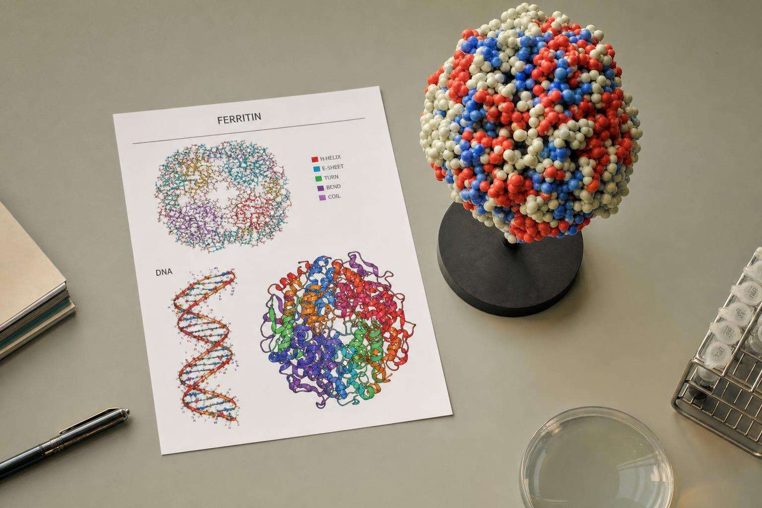
About 10–15% of inherited iron overload is non-HFE (HJV, HAMP, TFR2, ferroportin/SLC40A1). Juvenile hemochromatosis loads rapidly with early cardiac and endocrine disease. Negative HFE is not “not genetic”—escalate the panel when phenotype is severe.
Most hereditary hemochromatosis education stops at C282Y. The remaining slice is smaller but clinically louder when a young patient presents with heart failure and sky-high ferritin.
This article is informational and editorial only. It is not medical advice, diagnosis, or a treatment plan. Numbers and literature ranges cited here are not personal prescriptions. Consult a qualified clinician before changing medications, supplements, diet, equipment, or management of a diagnosed condition. Seek urgent care for emergencies.
How does AASLD classify non-HFE hereditary overload?
The AASLD 2011 guideline separates HFE-related disease from non-HFE inherited forms involving HJV, TFR2, ferroportin (SLC40A1), and HAMP, plus African iron overload and miscellaneous rare disorders. Roughly 10–15 percent of inherited overload after excluding C282Y/C282Y involves these non-HFE genes. C282Y homozygosity still dominates ordinary adult hereditary hemochromatosis practice.
EASL 2022 clinical practice guidelines update European investigation and management pathways for haemochromatosis including non-classical forms. When phenotype is classic or severe but HFE is wild-type, non-HFE genetic panels plus imaging or biopsy as needed are the rational next step—not reassurance that “genetics are negative so it is just lifestyle.”
What makes juvenile hemochromatosis different?
Juvenile disease, mainly HJV and rarely HAMP, suppresses the hepcidin pathway and packs iron into tissues over years rather than decades. Cardiac and endocrine presentations—cardiomyopathy, hypogonadism—appear in adolescence or early adulthood. That tempo is a genetic emergency of iron removal, not a wait-and-see ferritin hobby.
Do not stop the workup after a normal HFE panel in a young adult with cardiomyopathy and extreme iron studies. Specialty centers coordinate aggressive unloading, organ imaging, and family evaluation while the diagnosis is confirmed on expanded genetics.
| Gene | Pathway idea | Clinical note |
|---|---|---|
| HFE | Hepcidin regulation | Common adult type 1 HH |
| HJV / HAMP | Severe hepcidin failure | Juvenile, rapid, heart/endocrine |
| TFR2 | Hepatocyte sensing | AR, HFE-like parenchymal pattern |
| SLC40A1 loss | Export defect | Macrophage iron; often AD |
| SLC40A1 gain | Hepcidin resistance | Parenchymal-like pattern |
How does histology help sort primary patterns?
Hepatocyte-predominant iron suggests absorption-driven hepcidin-axis disease (HFE, TFR2, juvenile forms, gain-of-function ferroportin). Kupffer/macrophage-predominant iron suggests secondary overload or classic loss-of-function ferroportin disease. Pattern is a hypothesis generator, not a genotype. Biopsy still stages fibrosis when risk is high regardless of which gene is eventually named.
AASLD Liver Fellow Network teaching emphasizes mutations beyond HFE including HJV, HAMP, and TFR2 while reminding clinicians that C282Y homozygosity still dominates everyday hereditary hemochromatosis volumes.
What anti-patterns should teams avoid?
Stopping workup after negative HFE in a young patient with cardiomyopathy and high ferritin. Calling all high ferritin “type 1 hemochromatosis.” Confusing aceruloplasminemia with HFE disease. Assuming autosomal dominant ferroportin families follow only recessive sibling-screening scripts. Treating incomplete HFE panels as complete genetics.
For codified practice: escalate to non-HFE panels when phenotype is severe and HFE-negative; treat juvenile presentations as unloading emergencies; never use HFE-only testing as proof that iron overload is not genetic. See also EASL haemochromatosis guidance indexed on PubMed for contemporary European pathway language.
Editorial note: ranges and protocol bands cited here are literature and guideline context for shared decision-making with clinicians—not self-directed treatment schedules, home lab targets, or substitute care for emergencies or progressive organ disease.
Editorial note: ranges and protocol bands cited here are literature and guideline context for shared decision-making with clinicians—not self-directed treatment schedules, home lab targets, or substitute care for emergencies or progressive organ disease.
Editorial note: ranges and protocol bands cited here are literature and guideline context for shared decision-making with clinicians—not self-directed treatment schedules, home lab targets, or substitute care for emergencies or progressive organ disease.
Editorial note: ranges and protocol bands cited here are literature and guideline context for shared decision-making with clinicians—not self-directed treatment schedules, home lab targets, or substitute care for emergencies or progressive organ disease.
Editorial note: ranges and protocol bands cited here are literature and guideline context for shared decision-making with clinicians—not self-directed treatment schedules, home lab targets, or substitute care for emergencies or progressive organ disease.
Editorial note: ranges and protocol bands cited here are literature and guideline context for shared decision-making with clinicians—not self-directed treatment schedules, home lab targets, or substitute care for emergencies or progressive organ disease.
Editorial note: ranges and protocol bands cited here are literature and guideline context for shared decision-making with clinicians—not self-directed treatment schedules, home lab targets, or substitute care for emergencies or progressive organ disease.
Editorial note: ranges and protocol bands cited here are literature and guideline context for shared decision-making with clinicians—not self-directed treatment schedules, home lab targets, or substitute care for emergencies or progressive organ disease.
Editorial note: ranges and protocol bands cited here are literature and guideline context for shared decision-making with clinicians—not self-directed treatment schedules, home lab targets, or substitute care for emergencies or progressive organ disease.
Editorial note: ranges and protocol bands cited here are literature and guideline context for shared decision-making with clinicians—not self-directed treatment schedules, home lab targets, or substitute care for emergencies or progressive organ disease.
Sources & citations
Frequently asked