Metabolic Health
Iron Overload and Hemochromatosis: Labs, Genes, and Treatment
TSAT gates, ferritin myths, C282Y penetrance, phlebotomy targets, and why diet cannot replace blood removal.
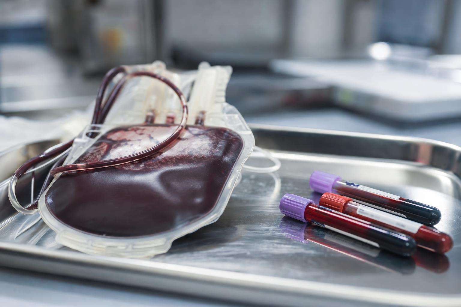
Classic hereditary hemochromatosis is most often HFE C282Y/C282Y-driven low hepcidin leading to excess intestinal iron absorption. Transferrin saturation ≥45% plus ferritin phenotype gates genetic testing; ferritin alone is not iron overload. Phlebotomy to ferritin about 50–100 µg/L is first-line for true overload—diet cannot remove multi-gram stores.
High ferritin is one of the most misread labs in primary care. Inflammation, alcohol, and fatty liver raise ferritin without iron overload, while true hereditary hemochromatosis can silently scar the liver before diagnosis. This guide locks genotype versus phenotype, sex differences, screening cascade logic, and therapy path separation between phlebotomy and chelation.
This article is informational and editorial only. It is not medical advice, diagnosis, or a treatment plan. Numbers and literature ranges cited here are not personal prescriptions. Consult a qualified clinician before changing medications, supplements, diet, or management of a diagnosed condition. Seek urgent care for emergencies.
What causes iron overload, and how is hereditary hemochromatosis different from high ferritin?
Iron overload means excess total body iron depositing in organs. Classic HFE hereditary hemochromatosis (HH) accounts for most northern European typical cases: about 80–85% of classic HH is C282Y/C282Y, which lowers hepcidin and increases gut iron absorption. Prevalence of C282Y homozygosity is roughly 1 in 220–250 among people of northern European ancestry. Other HFE genotypes (compound heterozygotes, H63D-related patterns) carry lower and more cofactor-dependent risk. Non-HFE genetic forms (juvenile HJV/HAMP, TFR2, ferroportin disease) are rarer and phenotypically distinct.
Secondary overload from transfusions, iron-loading anemias, or parenteral iron is a different mechanism—exogenous iron supply—often needing chelation rather than phlebotomy when anemia prevents bleeding therapy. Dysmetabolic or NAFLD-associated hyperferritinemia frequently shows high ferritin with normal or near-normal transferrin saturation and does not automatically equal multi-gram overload.
Penetrance is incomplete: biochemical expression of C282Y/C282Y is common (on the order of ~70% in many series) but severe organ disease is much less frequent (under ~10% in some framings; Australian HealthIron-type documented disease roughly 28% of men versus 1% of women in landmark reporting). About 30% of C282Y homozygotes in pooled screens can have normal ferritin. Genotype is not destiny.
| Pattern | Primary mechanism | TSAT pattern | First-line therapy direction |
|---|---|---|---|
| HFE C282Y/C282Y HH | Low hepcidin → ↑ absorption | Often ≥45% | Phlebotomy if overload confirmed |
| Milder HFE genotypes | Partial risk + cofactors | Variable | Phlebotomy only if true overload |
| Transfusional / secondary | Exogenous iron load | Context-dependent | Chelation when anemic / unbleedable |
| NAFLD / inflammatory hyperferritin | Metabolic/inflammation | Often normal | Treat liver/metabolic disease; not auto-bleed |
How is iron overload diagnosed—what thresholds actually matter?
Guideline-anchored phenotypic gating starts with transferrin saturation (TSAT) ≥45% plus ferritin interpretation. TSAT sensitivity for detecting C282Y/C282Y in classic algorithms is extremely high (reported about 97.9–100% in AASLD-era teaching). Ferritin reflects stores when inflammation is absent but rises as an acute-phase reactant—hence the primary-care trap.
Among C282Y homozygotes, ferritin >1000 µg/L marks substantially higher cirrhosis risk (series often cite roughly 20–45%), whereas ferritin under 1000 without other risk factors associates with advanced fibrosis rates under about 2% in classic teaching. That 1000 threshold is a risk stratifier, not a casual wellness number. HFE genotyping confirms classic HH after phenotypic suspicion; universal population genetic screening is not recommended by AASLD/ACG/EASL-class guidance. Cascade family screening after a proven proband is appropriate.
MRI liver iron concentration and specialist hepatology assessment refine organ iron and fibrosis staging when indicated. Liver biopsy is less first-line than in past decades but still has roles. Always separate stages: genetic susceptibility, biochemical overload, and organ damage.
Authoritative overviews for patients and clinicians include the NIDDK hemochromatosis pages and society guidelines (AASLD 2011 lineage; EASL 2022 updates on iron overload management).
What treatments work—and can diet replace phlebotomy?
Therapeutic phlebotomy is first-line for classic HFE overload in patients who can tolerate blood removal: often about 500 mL weekly or biweekly during induction until ferritin reaches roughly 50–100 µg/L, then maintenance tailored to keep stores low without frank iron deficiency. Early treatment before cirrhosis is the survival hinge; once cirrhosis exists, hepatocellular carcinoma surveillance continues even after iron depletion.
Diet is adjunctive, not curative. Daily dietary iron absorption differences (a few milligrams) cannot match removal of roughly 200–250 mg of iron per unit of blood. Practical advice usually includes avoiding iron and high-dose vitamin C supplements, avoiding raw shellfish (Vibrio risk in iron overload), and limiting alcohol especially with liver disease—not extreme iron-free diets that replace phlebotomy. Chelation is for secondary overload or when phlebotomy is impossible—not the default first path for classic ambulatory HFE HH.
Why do men and women present differently?
Men typically express biochemical and clinical disease earlier and more severely. Women often have partial protection from menstrual iron loss until menopause, after which risk converges upward. That sex difference explains some of the stark male-versus-female disease percentages in population studies and should shape counseling—not false reassurance that women cannot get HH. Pregnancy management of known HH requires specialist coordination; this article does not provide obstetric protocols.
Bottom line: high ferritin demands TSAT context; C282Y/C282Y explains most classic HH but penetrates incompletely; phlebotomy targets ferritin 50–100 µg/L; diet helps around the edges; cascade family testing yes, universal genetic screening no; and sex-tagged counseling prevents both panic and missed disease.
Sources & citations
Frequently asked