Metabolic Health
MRI Liver Iron, Cardiac T2*, and When Biopsy Still Matters
MRI quantifies liver iron concentration and cardiac T2* noninvasively. In HFE HH, biopsy is mainly for fibrosis staging when ferritin exceeds 1000 µg/L or enzymes rise—not routine genetic diagnosis.
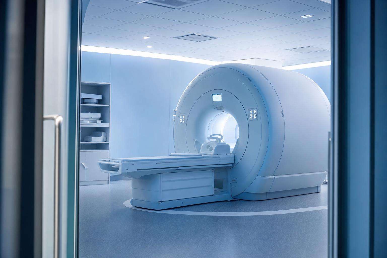
MRI quantifies liver iron concentration and, with cardiac T2*, myocardial iron—especially vital in transfusional overload. In classic HFE HH, biopsy is mainly for fibrosis staging when ferritin is >1000 µg/L or enzymes rise—not routine genetic diagnosis.
Iron workups used to mean automatic biopsy. Genetics and calibrated MRI reordered that sequence without making tissue staging obsolete when cirrhosis risk is high.
This article is informational and editorial only. It is not medical advice, diagnosis, or a treatment plan. Numbers and literature ranges cited here are not personal prescriptions. Consult a qualified clinician before changing medications, supplements, diet, equipment, or management of a diagnosed condition. Seek urgent care for emergencies.
How does MRI measure liver iron concentration?
Proton transverse relaxation methods (T2*, R2, R2*) shorten as liver iron rises. Validated sequences convert those signals into liver iron concentration estimates that correlate with biopsy hepatic iron content. The AASLD hemochromatosis guideline already noted good correlation between biopsy HIC and MRI relaxation methods; later radiology reviews describe MRI as state-of-the-art noninvasive LIC quantification for clinical management.
Example HIC bands in guideline tables place normal roughly around 5–27 µmol/g dry weight (about 300–1500 µg/g) and hereditary hemochromatosis often much higher (example ranges spanning tens to hundreds of micromoles per gram). One series noted all cirrhotics had HIC above about 200 µmol/g dry weight—roughly seven times the upper limit of normal in that framing.
MRI does not replace clinical judgment on hepatocellular carcinoma surveillance once cirrhosis is established. It does reduce needless needles when the only question was “how much iron is in the liver?”
When is liver biopsy still indicated?
After HFE testing, biopsy is less important for proving genetic hemochromatosis and more important for prognosis: advanced fibrosis and cirrhosis drive varices and HCC pathways. AASLD Rec 6: consider biopsy in C282Y homozygotes or compound heterozygotes if ALT/AST are elevated or ferritin exceeds 1000 µg/L.
When ferritin is under 1000 without other risks, advanced fibrosis/cirrhosis rates under about 2 percent are cited—biopsy often unnecessary. Dual pathology, non-HFE uncertainty, or discordant imaging still push toward tissue. If biopsying, stain Perls’ iron and stage fibrosis explicitly.
| Scenario | Preferred tool | Why |
|---|---|---|
| Classic HFE, ferritin <1000, normal enzymes | Noninvasive follow-up | Low advanced fibrosis probability |
| Ferritin >1000 or raised ALT/AST | Stage fibrosis (biopsy/elastography/MRI pathway) | Prognosis and surveillance |
| Transfusional overload | LIC MRI + cardiac T2* | Organ-specific iron maps therapy |
| Unclear non-HFE phenotype | Imaging ± biopsy | Pattern and dual disease |
Why is cardiac T2* essential in some patients?
Transfusional iron cardiomyopathy is a preventable disaster. Cardiac T2* falls as myocardial iron rises, giving a risk stratum that serum ferritin cannot fully replace. Multi-transfused patients need heart imaging even when “liver enzymes look fine.” Ignoring cardiac iron because the primary specialty is hepatology is a classic silo failure.
For classic adult HFE disease, cardiac iron is less often the lead problem than cirrhosis risk, but symptomatic or severe cases still deserve comprehensive organ assessment. Match imaging to mechanism: absorption-first HFE versus unit-after-unit siderosis.
What risks and anti-patterns surround biopsy and MRI?
Guideline-cited biopsy bleeding rates are roughly 1–6 percent with mortality under 1 in 10,000—non-zero, which is why non-invasive first is rational. Anti-patterns include universal biopsy for every C282Y with ferritin 400; treating historical hepatic iron index above 1.9 as a modern mandatory criterion; confusing steatosis MRI with iron quantification sequences; and claiming MRI alone cancels HCC surveillance after cirrhosis is proven.
Prefer non-invasive fibrosis assessment plus MRI LIC when available, then biopsy when dual pathology, non-HFE uncertainty, or high-risk ferritin/enzyme profiles demand tissue. CDC materials note MRI may be used to check for cirrhosis or scarring in care pathways—language that supports imaging without idolizing any single machine protocol.
See also Reeder and colleagues’ Radiology review for modern MRI LIC method context used by imaging services worldwide.
Editorial note: ranges and protocol bands cited here are literature and guideline context for shared decision-making with clinicians—not self-directed treatment schedules, home lab targets, or substitute care for emergencies or progressive organ disease.
Editorial note: ranges and protocol bands cited here are literature and guideline context for shared decision-making with clinicians—not self-directed treatment schedules, home lab targets, or substitute care for emergencies or progressive organ disease.
Editorial note: ranges and protocol bands cited here are literature and guideline context for shared decision-making with clinicians—not self-directed treatment schedules, home lab targets, or substitute care for emergencies or progressive organ disease.
Editorial note: ranges and protocol bands cited here are literature and guideline context for shared decision-making with clinicians—not self-directed treatment schedules, home lab targets, or substitute care for emergencies or progressive organ disease.
Editorial note: ranges and protocol bands cited here are literature and guideline context for shared decision-making with clinicians—not self-directed treatment schedules, home lab targets, or substitute care for emergencies or progressive organ disease.
Editorial note: ranges and protocol bands cited here are literature and guideline context for shared decision-making with clinicians—not self-directed treatment schedules, home lab targets, or substitute care for emergencies or progressive organ disease.
Sources & citations
Frequently asked